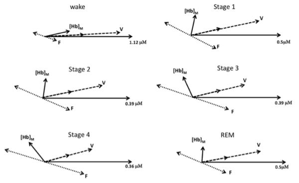
Fig. 7.
Phasor representation of the LFOs during various sleep conditions (wake, Stage1, Stage 2, Stage 3, Stage 4, REM). The numerical concentration quantifies the radial coordinate by specifying the amplitude of [HbO]M (the phasor of measured [HbO]) which is taken as the phase reference and thus is always represented by a horizontal phasor. The phasor of measured [Hb] is indicated by [Hb]M. The oxy-hemoglobin phasors associated with blood volume LFOs ([HbO]V) are indicated with V, while those associated with flow velocity ([HbO]F) are indicated with F. The corresponding deoxy-hemoglobin phasors are readily identified in the figure by recalling that [Hb]V = k[HbO]V and [Hb]F = −[HbO]F (see text for details).

