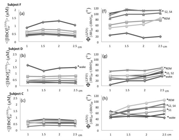
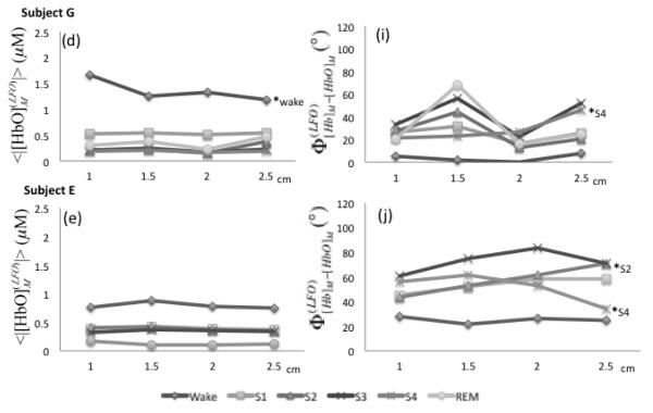
Fig. 9.
(a)-(e) Amplitudes of measured [HbO] LFOs as a function of source-detector distance in the range 1.0-2.5 cm (left side). (f)-(j) Phase difference between measured [Hb] and [HbO] LFOs. Sleep stages are indicated by different symbols. The shortest (1 cm) and farthest (2.5 cm) source-detector separation distances were compared within each sleep stage by t and U2 tests (p < 0.05 indicated by a * followed by the associated sleep stage).


