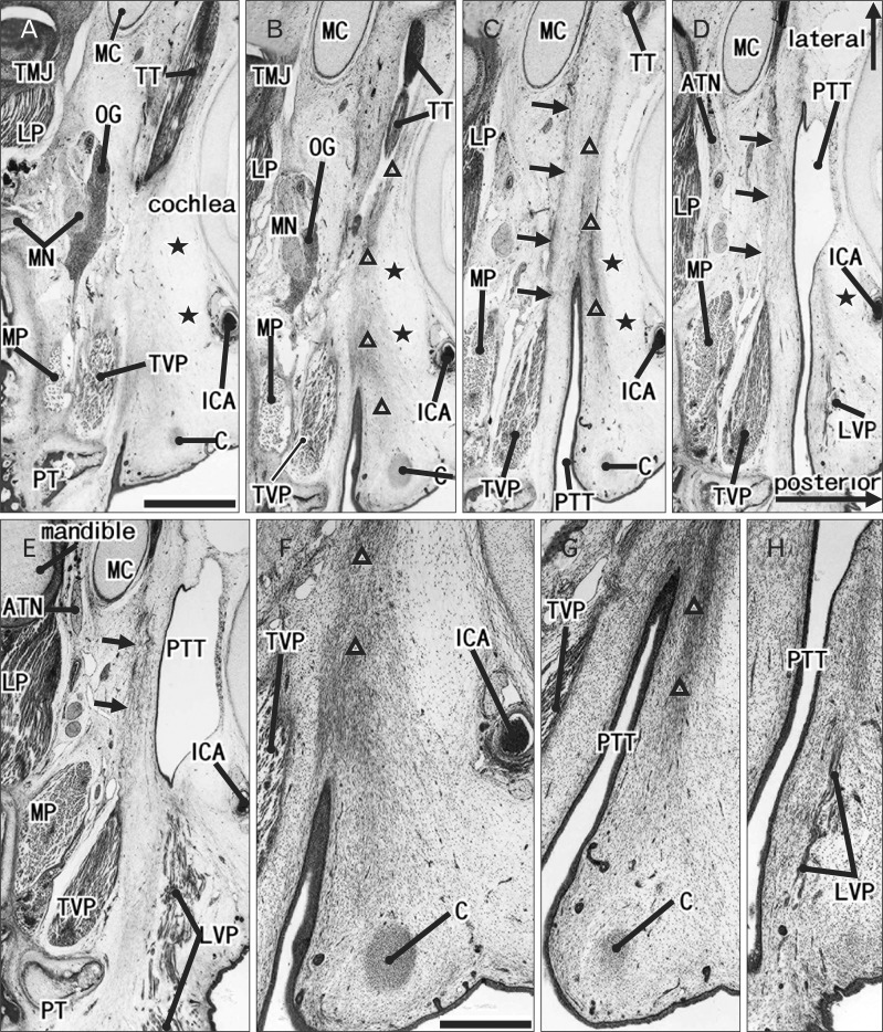
Fig. 1.
Initial stage of development of the pharyngotympanic tube cartilage: a fetus with 102 mm crown-rump length. Hematoxlyin and eosin staining. Horizontal sections. (A [E]) is the most superior (or inferior) side of the figure: the distance is 3 mm. The posterior or lateral orientation is shown by arrows in (D). (F-H) are higher-magnigfication views of the pharyngotympanic tube (PTT) shown in (B-D), respectively. (A [C]) displays the superior (or inferior) end of the tubal cartilage (C): thus, the initial cartilage, almost 1 mm in diameter, is restricted to the posterior side of the pharyngeal opening of the PTT. The superior end of the levator veli palatini muscle (LVP) is seen 1 mm below the cartilage (D). A thick, band-like mesenchymal condensation or an "adventitia" in the text (triangles) extends between the cartilage and the tensor tympani muscle (TT). A facia (arrows) connects between the tensor veli palatini muscle (TVP) and Meckel's cartilage (MC). The internal cartotid artery (ICA) does not run through the bony carotid canal but is exposed to a large loose space (stars) on the posterior side of the PTT. ATN, auriculotemporal nerve; LP, lateral pterygoid muscle; MN, mandibular nerver; MP, medial pterygoid muscle; OG, otic ganglion; PT, pterygoid process; TMJ, temporomandibular joint. Scale bars in (A)=1 mm (A-E); in (F)=0.5 mm (F-H).

