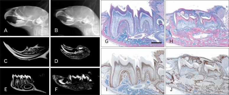
Fig. 2.
Disturbances in tooth formation and eruption failure of molars in Col1a1-cre:Catnb+/lox(ex3) mice. (A, B) In microradiography, general dimensions of craniofacial skeleton in MT mice are smaller than those in WT littermates. Particularly, mandible of MT is severely retarded and resulted in malocclusion. (C, D) In the mid-sagittal view, mandibular incisor is shorter and smaller than that of WT mice. (E, F) Molars of MT mice are impacted within mandible and root formation is impaired while those of WT mice are normally formed and erupted into the oral cavity. (G, H) In H&E-stained sagittal sections of the mandibles, bone deposition is remarkably increased and molars are not erupted in the MT mice. (I, J) In MT mice, β-catenin expression is upregulated in the odontoblasts and osteoblasts. MT, mutant; WT, wild type; H&E, hematoxylin and eosin. Scale bar=100 µm (G-J).

