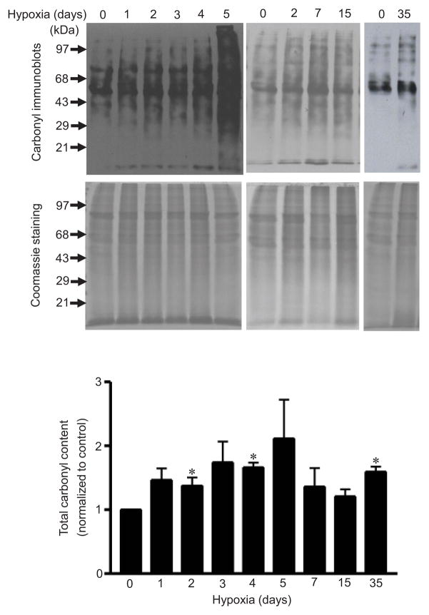
Fig. 3. Effects of chronic hypoxia on protein carbonylation in an in vivo model of pulmonary hypertension.
Rats were subjected to chronic hypoxia (10% O2) for indicated durations. After treatments, pulmonary arteries were isolated and homogenized. Proteins were derivatized with DNPH and subjected to Western blotting, with rabbit polyclonal IgG for DNP used to monitor carbonylated proteins. Total protein levels were monitored by Coomassie Blue staining. The bar graph represents means ± SEM (n = 3 – 4). *, P<0.05 vs. normoxia (0 day hypoxia).

