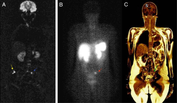
Figure 1 .
Comparison of DWI (A), 111 In-pentetreotide scintigraphy images (B) and T1-weighted (C) showing bone metastasis in left sacral bone detected by diffusion-weighted imaging (blue arrow) and confirmed by T1 sequence (green arrow). The hyperintense signal in the right subcutaneous fluid (yellow arrow) is a pitfall due to T2 shine-throught effect and should not be interpreted as metastasis. OctreoScan® image (B) reveals correlation between radiotracer uptake (red arrow) and MR findings.

