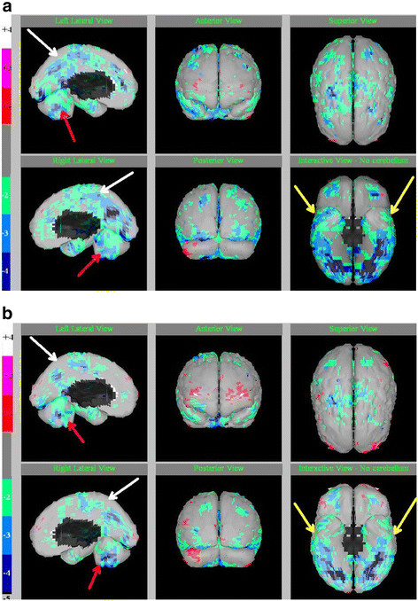
Figure 1.
SPECT scan images in a 12 year old boy with autism (a) before and (b) after 80 sessions of HBOT at 1.3 atm. Legend: minus 2 (green) to minus 4 (blue) standard deviations indicate the magnitude of regional hypofunctioning (hypoperfusion). White arrows indicate improvement in deeper cortical hypoperfusion patterns. Red arrows on sagittal slices show the midline cerebellum hypoperfusion and improvements after HBOT. Yellow arrows on the “underside” view show the temporal lobe hypoperfusion with improvements after HBOT. Pictures courtesy of J. Michael Uszler, MD. Credit: Permission for use of figure from Hyperbaric Oxygen for Neurological Disorders granted by Best Publishing Company, Palm Beach Gardens, FL.

