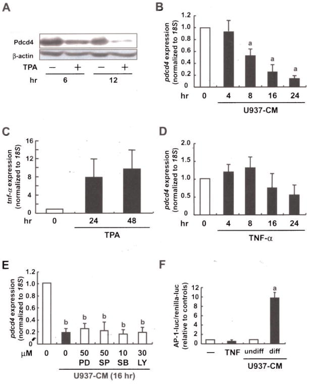
Figure 4.
Macrophage-released factors contribute to Pdcd4 regulation in tumor cells. (A) Shaved dorsal skin areas of 6-wk-old ICR mice were treated with the vehicle (acetone) or TPA (8 nmol). After 6 or 12 h, the mice were killed and the epidermis from each was collected, after which lysates from the epidermis samples were subjected to Western blot analysis (β-actin used as the control). (B) U937 cells were differentiated and activated with TPA (10 nM) for 48 h. Differentiated U937 cells were reseeded and allowed to condition medium for 24 h (U937-CM). Human MCF7 breast tumor cells were incubated with U937-CM for 4–24 h. (C) U937 cells were stimulated with TPA (10 nM) for 4, 24, or 48 h. (D) MCF7 cells were incubated with recombinant TNF-α (20 ng/mL) for 4–24 h. (E) MCF7 cells were preincubated with the indicated inhibitors for 30 min. After the preincubation, cells were treated with (C) U937-CM (as described above) or (D) TNF-α (20 ng/mL) for 24 h. For (B)–(E) total RNA was isolated and subjected to RT-qPCR (expression was normalized to 18S rRNA expression and given relative to control treatment). Student’s t-test was used to determine significant differences. aP <0.005 and bP <0.001 versus control. (F) MCF7 cells were cotransfected with AP-l luciferase construct and phRL-TK vector using Rotifect reagent. Transiently transfected cells were stimulated with TNF-α, or U937-CM (undifferentiated vs. differentiated) for 24 h. Luciferase activity was measured as described in the Materials and Methods Section. AP-1-luc activity was expressed relative to the respective controls. Student’s t-test was used to determine significant differences. aP <0.005 versus control.

