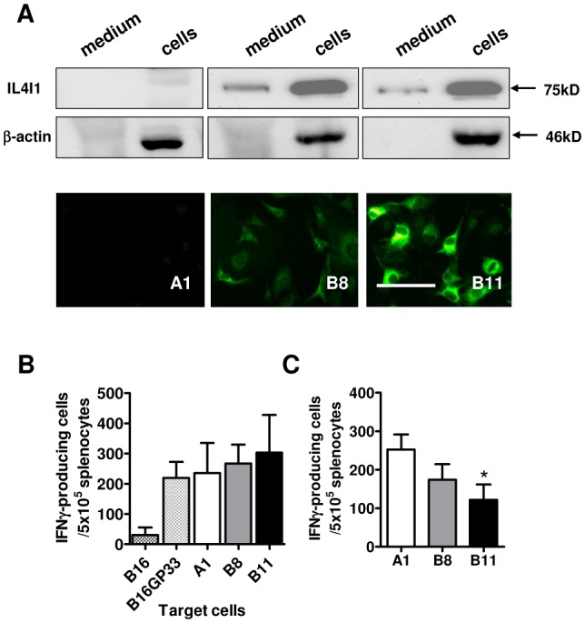
Figure 1. Characterization of IL4I1-expressing B16GP33 clones.

(A) Culture medium and whole cell lysate proteins from B16GP33 cells either transfected with an empty vector (A1) or with a vector coding for the myc-tagged murine IL4I1 protein (B8 and B11) were analyzed by Western blot (upper panel). IL4I1 was revealed by immunofluorescence (lower panel). Magnification ×400, scale bar = 100 μm. (B) ELISpot-IFNγ of anti-GP33 effectors against B16-derived cells. Splenocytes from GP33-vaccinated mice were cultured 24h with tumor cells, then the number of IFN-γ-producing cells was measured (mean from three experiments ±SD). (C) Ex-vivo ELISpot-IFNγ in tumor cell conditioned medium. Three-day conditioned media from 106 cells/ml tumor clones were used as culture medium for freshly isolated splenocytes from GP33-vaccinated mice. The number of IFN-γ-producing cells was measured after a 24h- incubation with GP33 (mean from six experiments ±SD; A1 vs B11, *p=0.020 p value of Mann-Withney test).
