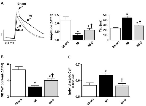
Figure 3. Effects of delapril on Ca2+ transients in LV myocytes.

A. Top left panel: Representative Ca2+transients from Sham (n=33), MI (n=27) and MI-D cardiomyocytes (n=41) loaded with Fluo-4 AM, observed by confocal microscopy. Top center and right panels: Average Ca2+-transient amplitude and Ca2+ reuptake kinetics (tau). B: SR Ca2+ content in Sham (n=9), MI (n=10) and MI-D cells (n=8) after caffeine application (100 μM). C. Mean ± S.E.M. of diastolic Ca2+ concentrations from Sham (n=23), MI cells (n=27) and MI-D cells (n=18) from the LV loaded with the fluorescent indicator Indo-1 AM. *P<0.05 in comparison with Sham mice. †P<0.05 in comparison with MI mice.
