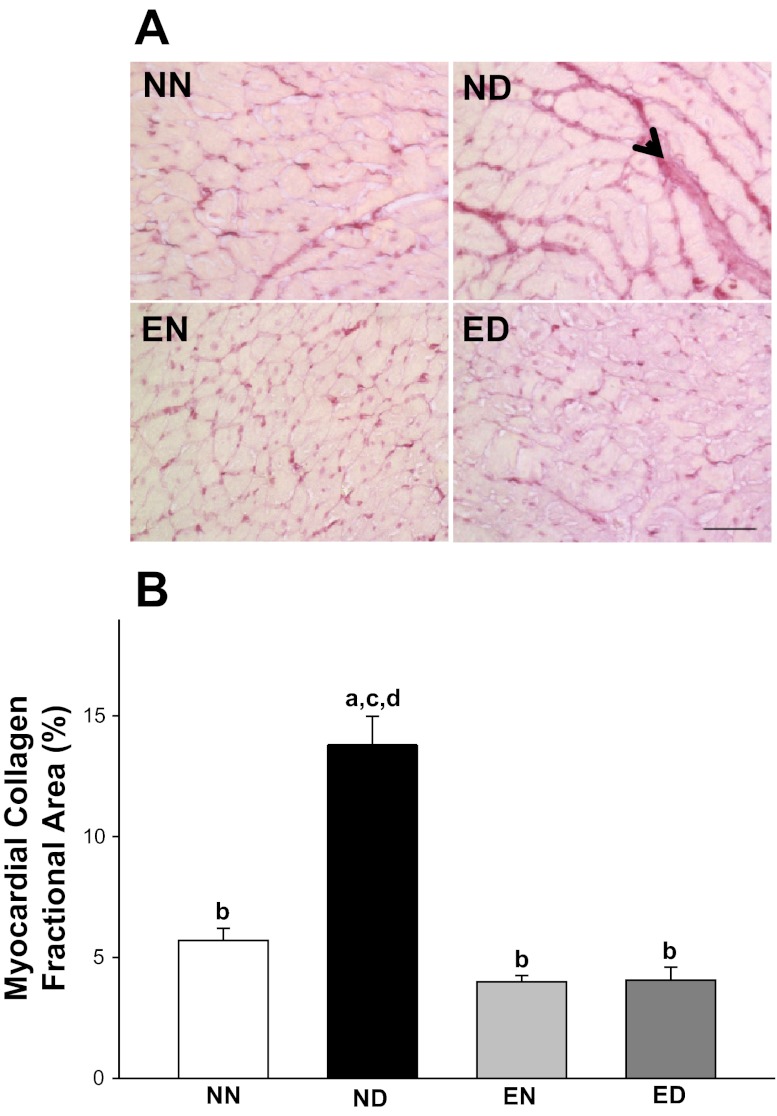
Fig. 3.
Exercise improved passive cardiac function in diabetes by preventing the accumulation of myocardial collagen. A: collagen-specific picrosirius red stained histological profiles of LV myocardium from all rat groups. The collagen distribution is highlighted by an arrowhead. Scale bar: 50 μm. B: LV myocardial collagen fractional area expressed as percent collagen fractional area in all four experimental groups. Values are means ± SE. Factorial (2 × 2) ANOVA interaction effect (health status × physical activity status) was significant (P = 0.001). Significant difference from a NN, b ND, c EN, and d ED: P ≤ 0.05.

