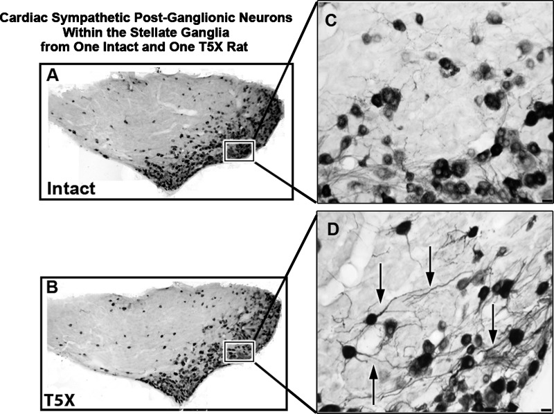
Fig. 5.
Cardiac sympathetic postganglionic neurons within the stellate ganglia from one intact and one T5X rat. Photomicrographs of a 30-μm horizontal section through the right stellate ganglia processed for cholera toxin B subunit immunoreactivity (black) from one sham-operated intact rat (A) and one T5X rat (B). CTB was injected into the pericardial sac to retrogradely label sympathetic postganglionic neurons projecting to the heart. The higher-power photomicrographs on the right (C and D) are of the boxed areas from A and B, showing details of CTB-labeled neurons and dendritic branching. Note the extensive dendritic branching (arrows) in the T5X rat compared with the intact rat. Bottom right scale bar = 25 μm in C and D.

