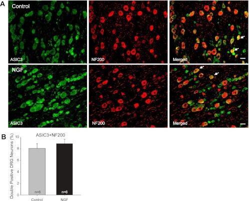
Fig. 7.
Colocalization of ASIC3 and NF200. Dual fluorescence immunohistochemistry was employed to examine labeling of ASIC3 and NF200. Note that NF200 was used to identify A-fiber of DRG neurons. A: photomicrographs are representative to illustrate staining of ASIC3 and NF200 in DRG neurons of a control limb (top panel) and a limb infused with NGF (bottom panel). Arrows indicate examples for merged ASIC3 and NF200 positive cells. There were no differences in the number of double staining of ASIC3 and NF200 in DRG neurons of control and infused groups. Scale bar = 50 μm. B: average data for colocalization of ASIC3 and NF-200, showing that there are no differences observed in the number of double staining for ASIC3 and NF200 in DRG neurons of control and NGF infusion groups. Note that no differences were observed in the number of NF200 positive DRG neurons in control and NGF infusion groups. One-way ANOVA was performed to compare variables for percentages of double-labeled neurons for ASIC3 and NF200.

