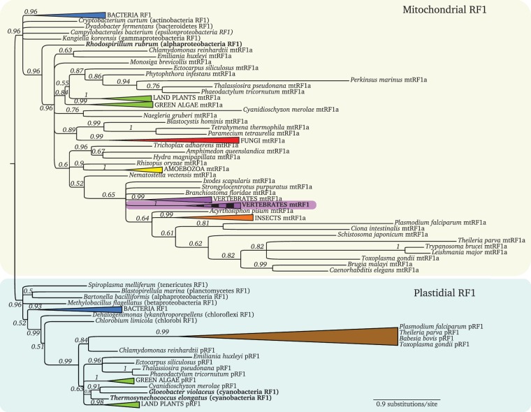
Fig. 2.
Bayesian RF1 phylogeny. The two main branches separate the mitochondrial proteins (yellow box) from the plastidial ones (green box). The mtRF1 branch nested within mtRF1a is highlighted in purple with a vertical striped pattern. Well-supported branches from well-established taxa were collapsed to improve readability (full noncollapsed tree in supplementary fig. 1, Supplementary Materials online). Alphaproteobacteria and cyanobacteria are highlighted in bold. (Colors for collapsed taxa: Blue—bacteria; green—Viridiplantae; red—fungi; yellow—amoebozoa; purple—vertebrates; orange—insects; and brown—apicomplexa.)

