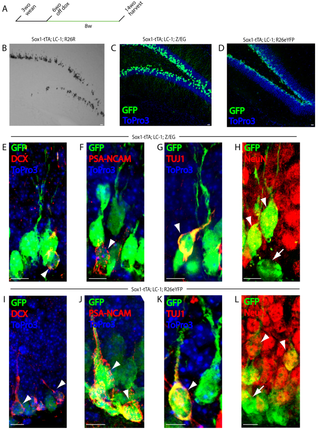
Fig. 3.
Sox1-expressing cells give rise to new neurons. (A) Schematic of the timeline for lineage-tracing experiments. Sox1-tTA; LC-1; reporter mice were taken off doxycycline at 6 weeks of age (system on) for 8 weeks then sacrificed at 14 weeks of age, except for R26R mice which were kept off doxycyline for 26 weeks. (B) X-gal staining of a 50 μm section of the dentate gyrus from an 8-month-old Sox1-tTA; LC-1; R26R mouse. (C) GFP (green) and DNA (ToPro3, blue) staining of a 50 μm section of the dentate gyrus from a 3.5-month-old Sox1-tTA; LC-1; Z/EG mouse. (D) GFP (green) and DNA (ToPro3, blue) staining of a 50 μm section of the dentate gyrus from a 3.5-month-old Sox1-tTA; LC-1; R26eYFP mouse. (E-H) GFP (green), DCX (red) and DNA (Topro3, blue) staining (E); GFP (green), PSA-NCAM (red) and DNA (Topro3, blue) staining (F); GFP (green), Tuj1 (red) and DNA (Topro3, blue) staining (G); and GFP (green) and NeuN (red) staining (H) of a 50 μm section of the dentate gyrus from a 3.5-month-old Sox1-tTA; LC-1; Z/EG mouse. (I-L) GFP (green), DCX (red) and DNA (Topro3, blue) staining (I); GFP (green), PSA-NCAM (red) and DNA (Topro3, blue) staining (J); GFP (green), Tuj1 (red) and DNA (Topro3, blue) staining (K); and GFP (green) and NeuN (red) (L) staining of a 50 μm section of the dentate gyrus from a 3.5-month-old Sox1-tTA; LC-1; R26eYFP mouse. All mice were taken off of doxycycline at 1.5 months. Arrowheads in E-L indicate cells that are double positive for GFP and described marker. Arrows in H and L indicate cells marked only by GFP. Scale bars: 10 μm.

