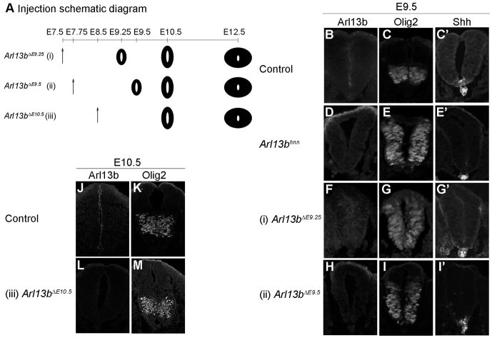
Fig. 2.
Distinct neural pattern when Arl13b is deleted at different time points. (A) Experimental design of tamoxifen injection to delete Arl13b. Tamoxifen is injected at E7.5 (i), E7.75 (ii) or E8.5 (iii), and embryos are collected at E9.5, E10.5 or E12.5 to examine neural tube patterning. Black neural tube indicates that ubiquitous Arl13b deletion has occurred, based on immunofluorescence with Arl13b antibody. (B,D,F,H) Arl13b expression can be observed in the ventricular zone of E9.5 control caudal neural tube (B), but not in E9.5 Arl13bhnn (D), Arl13bΔE9.25 (F) or Arl13bΔE9.5 (H). (C,E,G,I) Olig2 cells are specified in a restricted domain in E9.5 control (C), but are expanded in Arl13bhnn (E), Arl13bΔE9.25 (G) and Arl13bΔE9.5 (I) caudal neural tube. (C′,E′,G′,I′) Shh is expressed in the notochord and floor plate at E9.5 in both control and Arl13bΔE9.5 (C′,I′). Shh is only observed in the notochord in Arl13bhnn and Arl13bΔE9.25 (E′,G′). (J,L) Arl13b is expressed in the ventricular zone of E10.5 control caudal neural tube (J), but is absent in Arl13bΔE10.5 (L). (K,M) Olig2 cells are specified in their restricted domain in both control (K) and Arl13bΔE10.5 (M).

