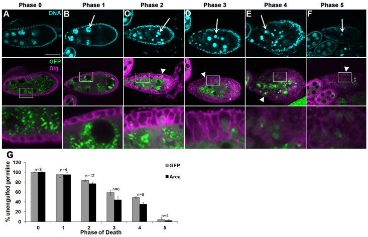
Fig. 1.
Progression of cell death and engulfment. Egg chambers from starved flies were labeled with DAPI to label DNA (cyan, top panels) and α-Dlg (magenta). Egg chambers express a germline-specific GFP gene trap (G89, green). Bottom panels are enlargements of boxed regions in middle panels. The state of chromatin condensation and fragmentation is used to characterize the different ‘phases’ of egg chamber degeneration. Egg chambers in Figs 1, 2, 3, 4, 5 and 6 are from stage 8 to early stage 9 of oogenesis. (A) Healthy (phase 0) egg chamber shows dispersed chromatin in nurse cell (NC) nuclei, and follicle cell (FC) nuclei surround the egg chamber. (B-F) Progression of cell death. NC nuclei become highly condensed and fragmented (arrows). Middle and bottom panels show that FC membranes enlarge and engulf germline GFP (arrowheads). (B) Phase 1: NC chromatin is disordered. (C) Phase 2: NC chromatin is condensed but individual nuclear regions are still apparent. (D) Phase 3: NC chromatin becomes highly condensed into individual balls. (E) Phase 4: NC chromatin is fragmented and widely dispersed. (F) Phase 5: few NC nuclear fragments remain. Scale bar: 50 μm in top and middle rows. (G) Quantification of engulfment from control G71/+ and GR1-GAL4 G89/TM6B flies shows decrease in germline GFP and area as death progresses. Data are mean±s.e.m.

