Fig. 2.
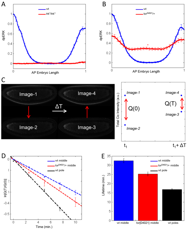
Cic lifetime is reduced by Torso signaling. (A) Quantified average dpERK gradients in the control (Histone GFP, blue) and the loss-of-function mutant trk1/trk1 (red) Drosophila embryos. The dpERK levels in the main body regions of the wild-type embryos are the same as in the trk1/trk1 embryos. Thus, the main body region of the wild-type embryos corresponds to zero level of Torso activation. Error bars correspond to s.e.m. Numbers of embryos used in the analysis: nwt=21 and ntrk1/trk1=17. (B) Quantified average dpERK gradients in the control (blue) and gain-of-function mutant torD4021/+ (red). The levels in the central region of mutant embryos are significantly increased. nwt=24 and ntorD4021=24. (C) Outline of the optical pulse labeling experiment. A population of Dronpa-tagged molecules is converted to the dark state at time t1. After a waiting time ΔT, the dark-converted population is converted back to the bright state. The fraction of surviving Cic-Dronpa, Q(T)/Q(0) is a measure of its lifetime. (D) First-order fits to survival kinetics of Cic-Dronpa in the middle of wild-type embryos (blue), in the middle of torD4021/+ embryos (red) and in the pole region of wild-type embryos (black). Error bars are s.e.m., based on a minimum of 40 samples for each waiting time. (E) Cic lifetimes at three different levels of RTK activation. Error bars correspond to 95% confidence intervals.

