Fig. 3.
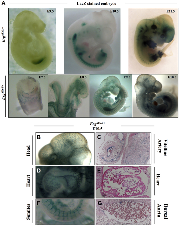
lacZ expression pattern during development of ErgΔEx3/+ and ErgΔEx4/+ mouse embryos. (A) lacZ staining corresponding to ErgEx3 was observed in the somites and limbs corresponding to developing articular cartilage starting at E9.5. By contrast, ErgEx4 expression was detected in the blood islands at E7.5 and then in all embryonic vasculature. (B,D,F) lacZ staining of whole-mount preparations of the head (B), heart (D) and somites (F) from E10.5 embryos displaying a clear endothelial restricted pattern in the brain vasculature, endocardium and intersomitic vessels, respectively. (C,E,G) lacZ staining of sectioned E10.5 embryos. lacZ-positive cells are seen in the vitelline artery (C), endocardium of the heart (E) and in ECs lining the dorsal aorta (G).

