
Fig. 5.
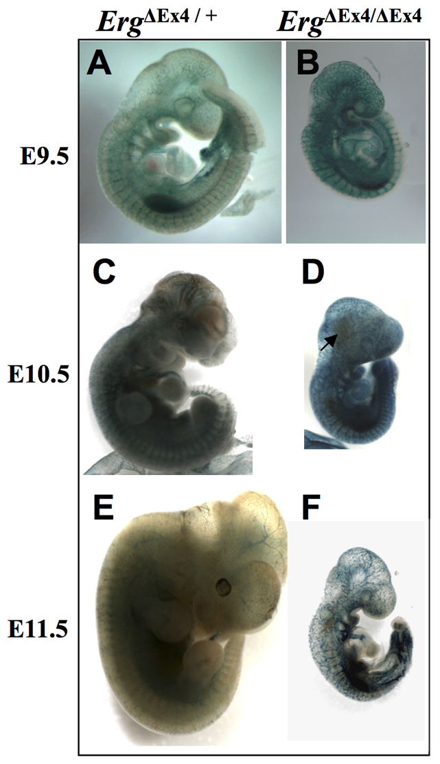
Morphology of ErgΔEx4/+ and ErgΔEx4/ΔEx4 embryos. (A,B) At E9.5, lacZ expression in ErgΔEx4/+ is seen in all embryonic vasculature. The ErgΔEx4/ΔEx4 embryos were smaller (B), but did not display any other significant morphological differences when compared with controls (A). (C,D) By E10.5, ErgΔEx4/ΔEx4 embryos (D) were notably smaller than control littermates (C), were paler and displayed disrupted vessels associated with hematoma (arrows). (E,F) Surviving ErgΔEx4/ΔEx4 embryos at E11.5 (F) showed a dramatic reduction in size and development compared with controls (E).