
Fig. 6.
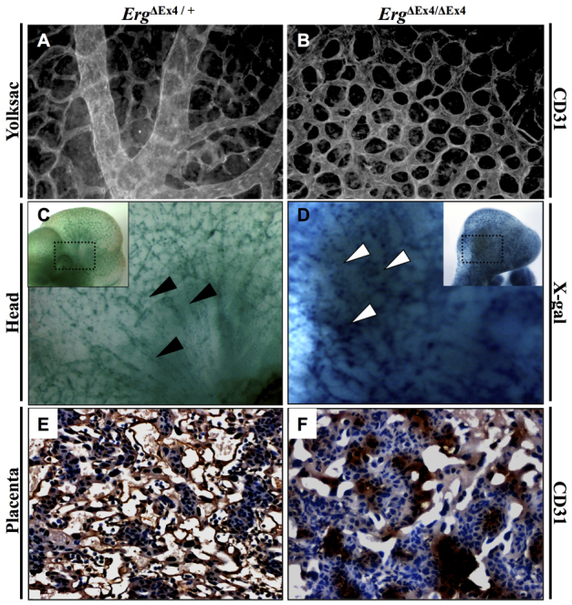
Vascular remodeling defects seen in ErgΔEx4/ΔEx4 embryos at E10.5. (A,B) CD31 staining shows that mutant yolk sac (B) lacks larger vessels, displaying only a capillary plexus when compared with controls that display a well-organized vasculature (A). (C,D) ErgΔEx4/ΔEx4 embryos show vascular remodeling abnormalities in the head (D), failing to exhibit normal alignment and invasion of vessels from the perineural vascular plexus into the developing ganglionic eminence, as seen in controls (C) (arrowheads). Insets are low-magnification images of the head. (E,F) CD31 staining of vasculature in the placental labyrinth at E10.5 shows abundant vessels in ErgΔEx4/+ (E), whereas the ErgΔEx4/ΔEx4 embryos display collapsed and pooled vessels (F).