
Fig. 7.
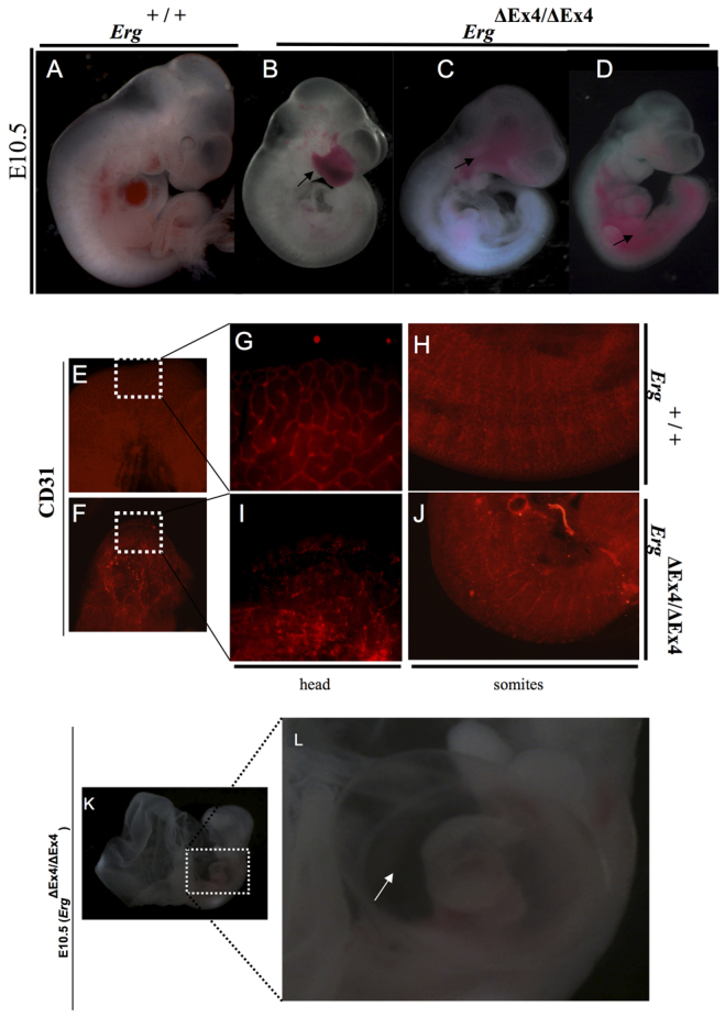
ErgΔEx4/ΔEx4 embryos display hemorrhages and disrupted vasculature. (A-D) Whole-mount images of Erg+/+ and ErgΔEx4/ΔEx4 mouse embryos at E10.5. ErgΔEx4/ΔEx4 embryos are growth retarded (B-D) and show hemorrhages (arrows in B-D) when compared with controls (A). (E,F) CD31 whole-mount immunohistochemistry of head of Erg+/+ (E) and ErgΔEx4/ΔEx4 (F) embryos at E10.5. (G-J) Magnified images of the boxed regions in E and F. Note the well-connected minor branches in the controls (G), whereas the mutants display a disorganized pattern of vasculature (H). (I) Wild-type embryo showing a segmented pattern of intersomitic vessels and capillary network, as compared with the severe disorganization of intersomitic vessels and reduced capillary network in ErgΔEx4/ΔEx4 embryos (J). (K,L) E10.5 ErgΔEx4/ΔEx4 embryo revealing pericardial effusion (K); the boxed region is magnified in L (arrow points to the effusion).