
Fig. 8.
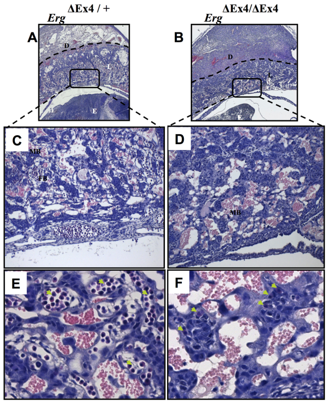
Placental vascular defects in ErgΔEx4/ΔEx4 embryos at E10. (A,B) The dashed lines in Erg+/+ (A) and ErgΔEx4/ΔEx4 (B) indicate the extent of decidual invasion in each placental section. (C,D) Magnification of the boxed areas in A and B. Well-defined vasculature is observed in Erg+/+ placentas (C), whereas the mutants, although they have abundant maternal blood sinuses, lack well-defined fetal-derived capillaries (D). (E,F) Higher magnification of C and D reveal abundant nucleated fetal blood in the controls (arrows, E), whereas the mutants show a drastic reduction in the number of fetal blood cells and exhibit collapsed capillaries (arrows, F). D, maternal decidua; L, labyrinth; E, embryo; MB, maternal blood; EB, embryonic blood.