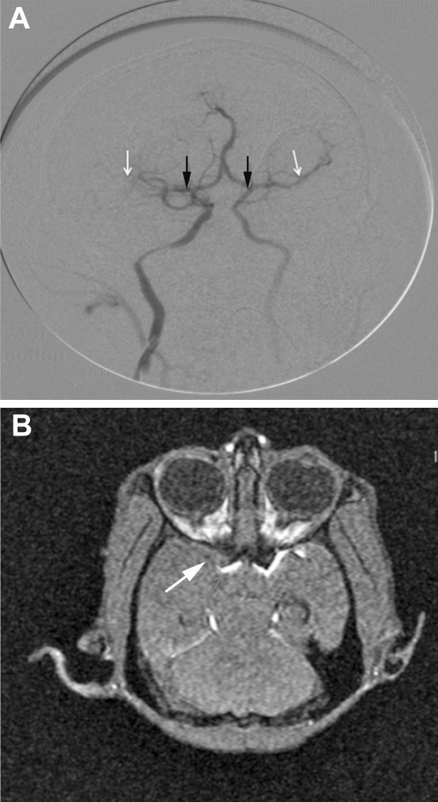
Figure 3.
After MCAO, contrast material was injected into the vasculature via the femoral catheter, and the cerebral arteries were visualized by using a C-arm fluoroscopy system. (A) The MCA (white arrows) is blocked on the ipsilateral side (black arrows indicate the origin of the MCA). (B) MCAO. The arrow and regions of white hyperintensity indicate blood flow in the MCA.

