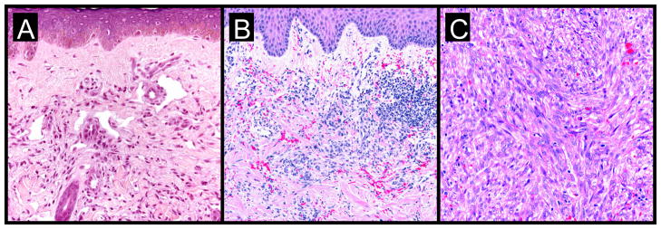
Figure 2. KS progression: a histological view.
- subtle proliferation of irregular vascular channels between normal stromal collagen
- the extravasation of erythrocytes and hemosiderin into the stroma
- detection of lymphoplasmacytic infiltrate
- proliferating spindled cells that form interlacing bundles closely approximated with blood-filled vascular spaces
- intracellular hyaline globules within lesional cells (likely representing phagocytosed erythrocytes within lysozomes)
- increased inflammatory infiltrate consisting of lymphocytes, plasma cells, macrophages, and dendritic cells
- formation of intersecting fascicles and sheets of proliferating spindled cells

