
Figure 7.
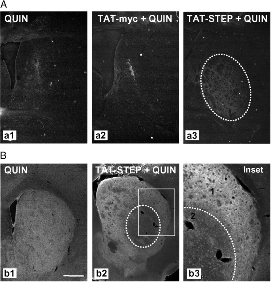
Intrastriatal TAT–STEP injection increases QUIN-induced cell death in R6/1 mice striatum. Control peptide (TAT–myc) or TAT–STEP was intrastriatally injected in R6/1 mice 1 h before intrastriatal QUIN injection. A, Cell death was assessed by Fluoro-Jade staining 24 h after QUIN injection. Representative photomicrographs show the striatal area occupied by Fluoro-Jade-positive cells in R6/1 mice striatum injected with QUIN (a1), TAT–myc plus QUIN (a2), or TAT–STEP plus QUIN (a3). B, Immunohistochemical staining against pERK2 was performed 24 h after QUIN injection in R6/1 mice striatum with or without previous injection of TAT–STEP. Representative images showing the striatum in all the conditions analyzed. In the inset, note the distinct pERK2 immunoreactivity in non-injured (1) and injured (2) striatal cells. Scale bar, 500 μm.