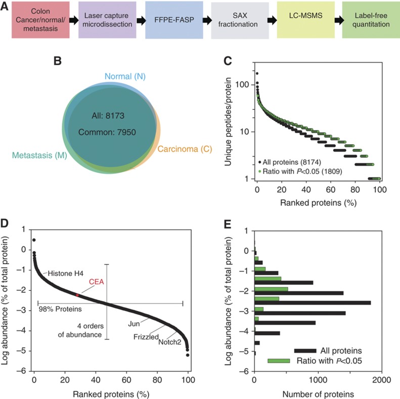
Figure 1.
Proteomic analysis of FFPE archival samples of colonic mucosa, cancer, and metastasis. (A) Proteomic workflow applied to microdissected samples of colonic mucosa (N), cancer (C), and metastasis (M). (B) Overlap of the proteins identified from colonic mucosa, cancer, and metastasis. (C) Peptide-based identification of proteins. (D) Distribution of protein abundances with selected examples. In red, relative abundance of CEA. Examples of lower abundant proteins identified in this study: Jun, proto-oncogene Jun; Frizzled, a G protein-coupled receptor protein that serves as receptor in the Wnt signaling pathway; and Notch 2. (E) Comparison of abundance distribution of all proteins and proteins that were significantly changed in adenocarcinomas. The protein abundances were calculated on the basis of total peptide intensities of all the quantified proteins.

