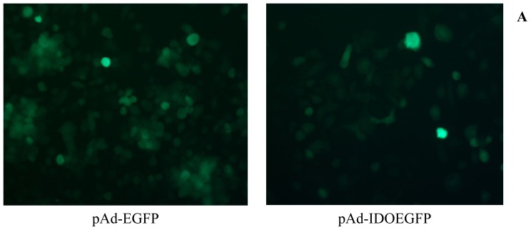
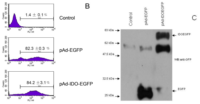
Figure 1.
The expression of IDOEGFP in MT-2 cells. MT-2 cells were infected with either pAd-EGFP or pAd-IDOEGFP at MOI of 100 for 60 h. (A) After 60 h of infection, the infection was monitored by EGFP expression under fluorescent microscopy. Original magnification 300×; (B) The efficiency of infection was determined by flow cytometry. After 60 h of infection, the cells were harvested and the numbers of EGFP positive cells were estimated by flow cytometry. Data are shown as mean ± SD, representative of three independent experiments; (C) Western blot analysis of IDOEGFP expression. After 60 h of infection, the noninfected (control) and infected cells were harvested and cell lysates from about 3 × 10⁵ cells were fractionated by SDS-PAGE. IDOEGFP protein was detected using purified mouse monoclonal anti-GFP antibody at a concentration of 1:1000. EGFP = enhanced green fluorescent protein; IDO = indoleamine 2,3-dioxygenase; MOI = multiplicity of infection; pAd-EGFP = recombinant adenovirus containing EGFP gene; pAd-IDOEGFP = recombinant adenovirus containing IDOEGFP gene; SDS-PAGE = sodium dodecyl sulfate-polyacrylamide gel electrophoresis.


