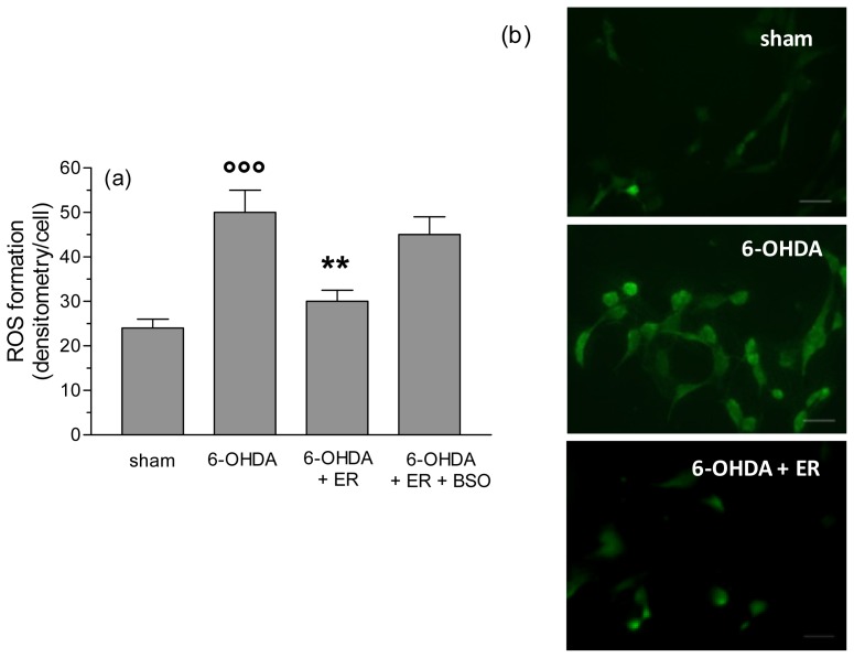
Figure 4.
ER counteracts 6-OHDA-induced intracellular ROS formation in SH-SY5Y cells. (a) SH-SY5Y cells were incubated with ER (5 μmol/L) in absence or presence of BSO (400 μmol/L) for 24 h and then treated with 6-OHDA (200 μmol/L) for 2 h. At the end of incubation, ROS formation was determined using a fluorescence probe, DCFH-DA, as described in the Experimental Section. Four randomly selected areas with 50–100 cells in each were analyzed under a fluorescence microscope and the values obtained are expressed as densitometry/cell. Values are shown as mean ± SEM of four independent experiments: ∘∘∘ p < 0.001 versus untreated cells, ** p < 0.01 versus cells treated with 6-OHDA at ANOVA with Bonferroni post hoc test; (b) representative images of ROS formation. Scale bars: 100 μm.

