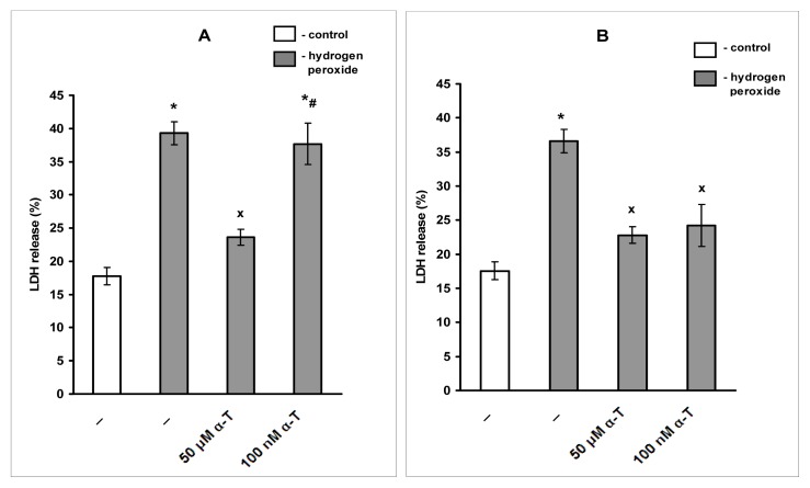
Figure 6.
The figure shows the effect of pre-incubation of immature cortical neurons with nanomolar and micromolar α-T for 0.5 and 18 h on the viability of these cells exposed to hydrogen peroxide. The results of one typical experiment (n = 6) are presented as means ± SEM of 3–4 parallel determinations. The neurons were isolated from the brain cortex of an embryonic rat brain as described under Experimental procedure. After 6 days in culture (at the 7th day in vitro) immature cortical neurons were pre-incubated for 0.5 h (A) or 18 h (B) with α-T and then exposed to 0.2 mM H2O2 for 24 h. The differences are significant by one-way ANOVA followed by Tukey’s multiple comparison test: * as compared to control values, p < 0.01; x as compared to the effect of H2O2 alone, p < 0.01; # as compared to the effect of H2O2 and 50 μM α-T, p < 0.05.

