Figure 1.
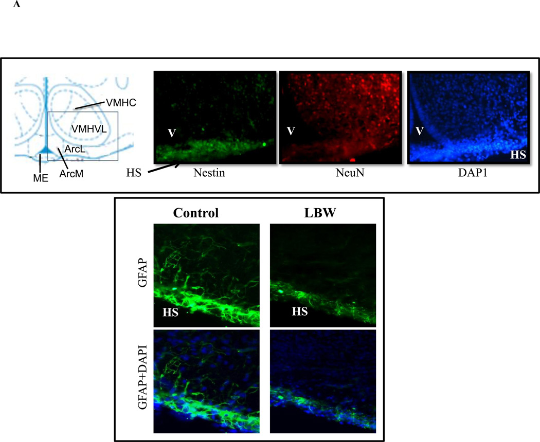
A: Evidence of Hypothalamic NPCs and GFAP in 1 Day Newborn
Brain from 1 day newborn was fixed, sectioned (20µm) and stained for markers of NPC (nestin), neuronal (NeuN), astrocyte (GFAP) and nuclei (DAPI). Upper images show evidence of hypothalamic NPCs around third venticular (V) region and lower images show hypothalamic surface (HS) GFAP (astrocyte). Images shown are at ×40 magnification.
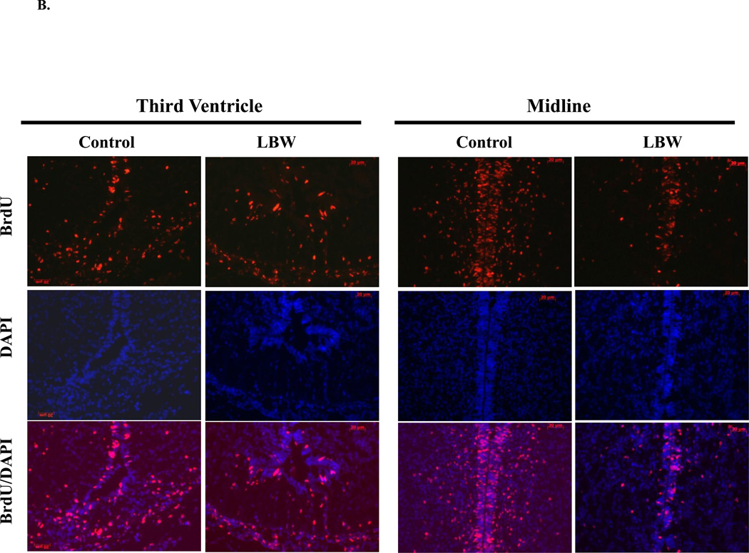
B: In Vivo NPC Proliferation and Migration: Hypothalamic Immunostaining of BrdU Incorporation
Food-restricted (n=3) and control (n=3) pregnant dams were injected with BrdU (50 mg/kg/day, i.p.) from e17–e19. After birth, brains were collected from 1 day old Control ( ) and LBW (
 ) newborn males. The images (×20) show hypothalamic BrdU (cell proliferation) and DAPI (nuclear marker) immunostaining around third venticular (V) region.
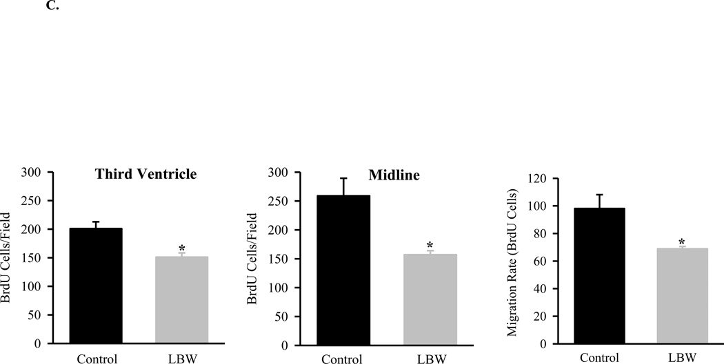
C: In Vivo NPC Proliferation and Migration
Food-restricted (n=3) and control (n=3) pregnant dams were injected with BrdU (50 mg/kg/day, i.p.) from e17–e19. After birth, brains were collected from 1 day old Control ( ) and LBW (
 ) newborn males. Three brains per litter were frozen, and three sections per brain were immunostained. Cell proliferation was determined by counting BrdU positive cells in third ventricle and midline. Migration rate was determined by counting BrdU labeled cells in the area between 30 µm to 100 µm from midline. The average of BrdU-labeled cell numbers of three sections represented one brain and average of three brain cell numbers represented one litter. Values are mean±SE; *P<0.05 vs. Control.


