Figure 2.
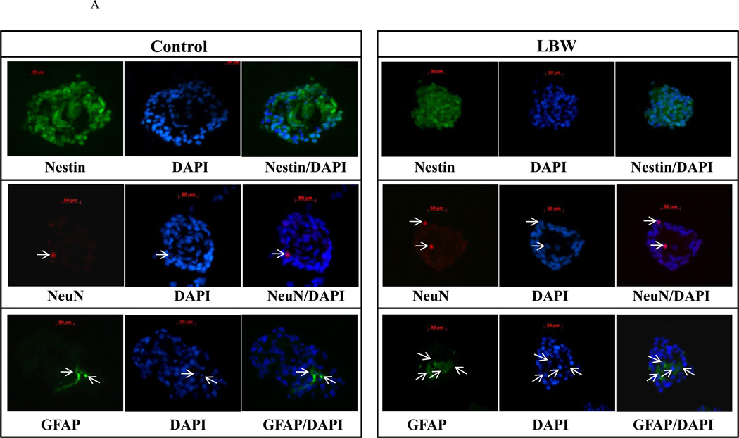
A: Nestin immunostaining of Neurosphere Sections
Hypothalamic NPC from 1 day old Control ( ) and LBW (
 ) newborn males were cultured in complete media. Neurospheres were sectioned (5 µm) and immunostained with nestin, Neu and GFAP. Images of LBW and Control are shown at 40× magnification.
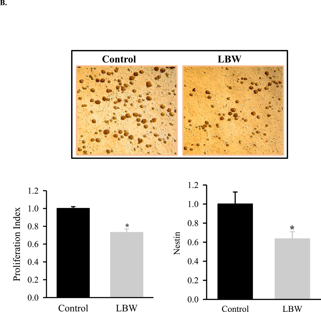
B: Basal Hypothalamic NPC Proliferation
Hypothalamic NPC from 1 day old Control ( ) and LBW (
 ) newborn males were cultured in complete media. Live images (magnification ×20), basal cell proliferation rate and nestin protein expression of LBW and Control NPCs. Values are fold change (mean ± SE); * P < 0.05 LBW vs. Control.
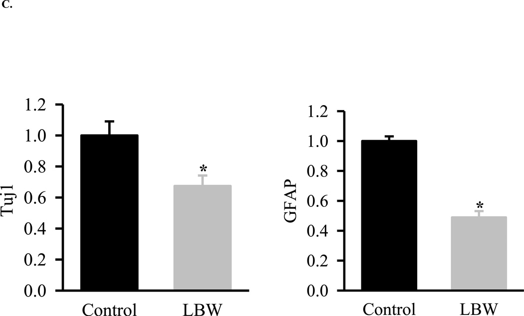
C: Basal Hypothalamic NPC Differentiation
Hypothalamic NPC from 1 Control ( ) and LBW (
 ) newborn males were cultured in differentiating media. NPC were harvested and protein expression of neuronal marker (Tuj1)) and astrocyte marker (GFAP) were determined. Values are fold change (mean ± SE); * P < 0.05 LBW vs. Control.


