Figure 3.
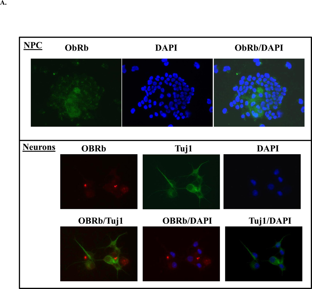
A: Evidence for NPC and Neuronal Leptin Receptor
NPC grown in complete or differentiating media were immunostained for leptin receptor (ObRb), neuronal marker (Tuj1) and nuclear stain (DAPI).
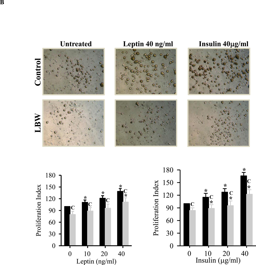
B: Leptin and Insulin Stimulated Hypothalamic NPC Proliferation
Hypothalamic NPC from 1 day old Control ( ) and LBW (
 ) males were cultured in complete media. On second day of seeding, NS were treated with leptin/insulin every 48h for 8 days and cell proliferation rate was determined. Values are percentage of untreated Control (mean ± SE); * P < 0.05 vs. untreated NS; “C” P < 0.05 LBW vs. Control.
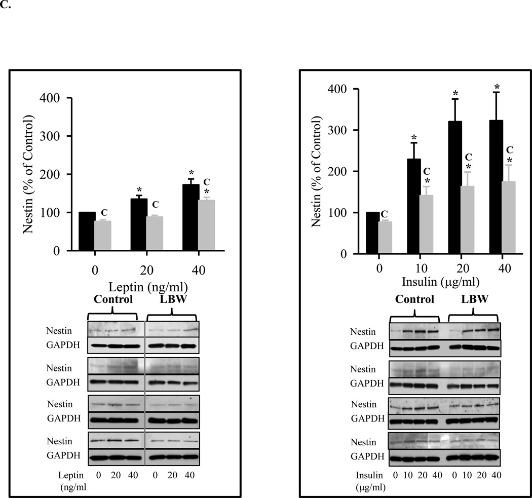
C: Leptin and Insulin Stimulated Hypothalamic Nestin Expression
Hypothalamic NS from 1 day old Control ( ) and LBW (
 ) males were cultured in complete media. On second day of seeding, NS were treated with leptin/insulin every 48h for 8 days. Nestin protein expression (marker of NPC) was determined. Values are mean ± SE; * P < 0.05 vs. untreated NS; “C” P < 0.05 LBW vs. Control.


