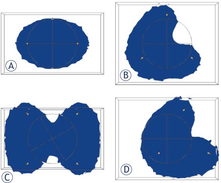
FIGURE 3.
Cross section of the tumor (circle) and its ablation zone (blue) at an applied voltage of 5.05 kV, which corresponds to the VIRE for two electrodes at a distance of 2.5 cm and depth of 1 cm for A) two electrodes B) three electrodes C) four electrodes D) three electrodes with the center of the electrodes shifted to the right 1 cm Note the asymmetry in B and the narrowing of the ablation zone in C. A bounding box around the tumor was used in the FEM simulations to improve the quality of the meshing and computations.

