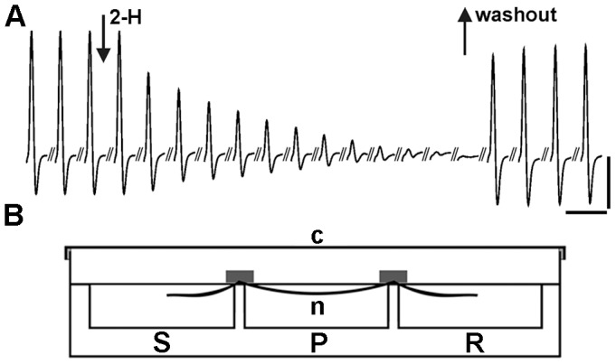
Figure 5. The evoked nCAP from the isolated rat sciatic nerve exposed to saline and 2-H.

. a) The amplitude of the nCAP, baseline to peak, was used as the main parameter to quantify the vitality of the sciatic nerve fibres during exposure to 2-H. The record presents the decrease in the amplitude nCAP during exposure of the sciatic nerve to 3.41 mg/mL 2-H. The first arrow indicates the beginning of the application and the second arrow indicates the time when the nerve was washed and bathed in normal saline. During exposure to 2-H, records were taken at a rate of 1 nCAP per 20s. After 2-H was replaced with normal saline, measurements were taken every 15 min. Vertical scale bar: 3 mV. Horizontal scale bar: 6 ms. b). Diagrammatic representation of the three-chamber recording bath made of Plexiglass. It consists of the recording (R), the perfusion (P) and the stimulating chambers (S), separated by two partitions. The sciatic nerve was placed along the three chambers which were filled with oxygenated saline to cover the nerve. The dimensions of each chamber were 26×26×10 mm (length. width, depth), total volume 10 mL. The cover (c) made of Plexiglass was used to close hermitically the whole recording system, while the air inside the bath was saturated with 2-H, to eliminate the evaporation of 2-H in the perfusion chamber.
