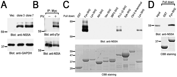
Figure 1. Identification of HCV NS5A-interacting proteins in B cells.
(A) Generation of stable B cell lines expressing HCV NS5A. Detergent soluble cell lysates from vector cells (Vec) and Myc-His-NS5A expressing clones (clones 3 and 7) were separated by SDS-PAGE and analyzed with immunoblotting with anti-NS5A and anti-GAPDH mAbs. (B) BJAB cells expressing Myc-His-NS5A (clone 7) were treated without (−) or with (+) PV and solubilized in the lysis buffer. Cell lysates were immunoprecipitated with anti-Myc mAb and immunoprecipitated proteins were separated by SDS-PAGE and analyzed with immunoblotting with anti-pTyr (PY20) and anti-NS5A mAbs. PV-treated cells expressing Myc-His-NS5A (clone 7) (C) or Huh-7.5 cells stably harboring an HCV subgenomic replicon (D) were solubilized in the binding buffer. Precleared lysates were reacted with the indicated GST-fusion proteins and binding proteins were separated by SDS-PAGE and analyzed with immunoblotting with anti-NS5A mAb. The amount of GST-fusion proteins was confirmed by Coomassie brilliant blue (CBB) staining (C and D). Molecular sizing markers are indicated at left in kilodalton. The results were representative of three independent experiments. Similar results were obtained when another line was examined (B and C).

