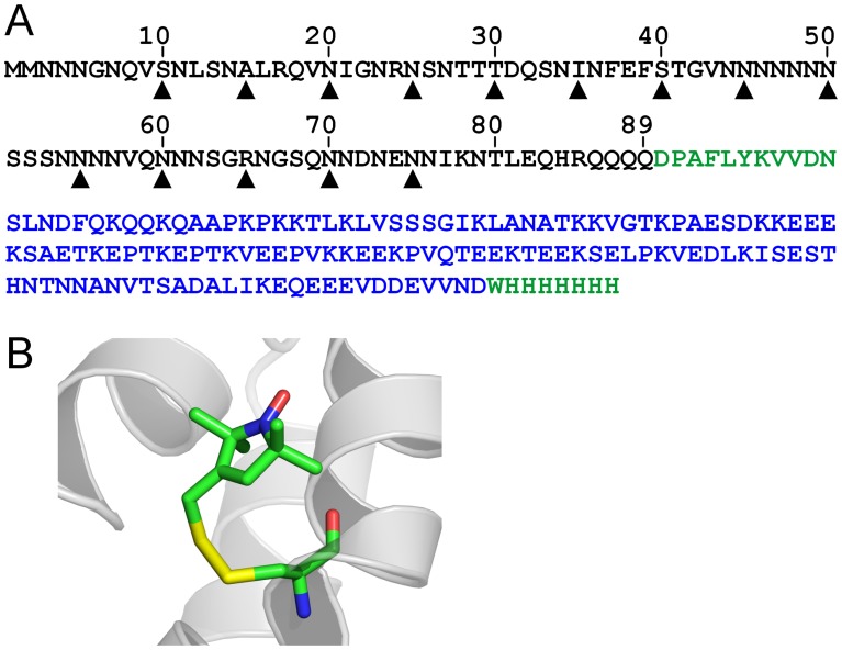
Figure 1. Spin labeling of Ure2p1–89-M.
(A) Sequence of Ure2p prion domain construct with positions for spin labeling indicated by arrow heads. The amino acid sequences of Ure2p prion domain, Sup35p M domain, and other linker/tag regions are shown in black, blue, and green, respectively. (B) A stick model of spin label R1 in the crystal structure of spin-labeled T4 lysozyme (PDB entry 2IGC).

