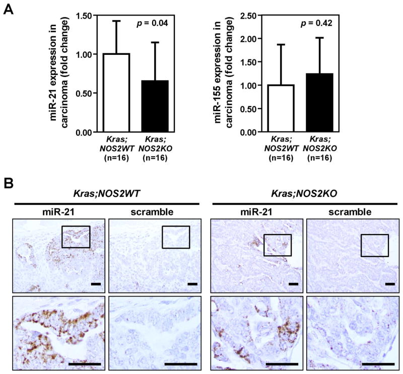
Figure 4.
Reduced miR-21 expression in lung carcinomas from KRASG12D;NOS2KO mice. A, MicroRNA expression in lung adenocarcinomas from KRASG12D;NOS2WT and KRASG12D;NOS2KO mice by quantitative RT-PCR analysis (mean ± SD). Carcinoma areas from KRASG12D;NOS2WT and KRASG12D;NOS2KO mice were macroscopically dissected from parafin-embedded lung sections for RNA isolation. P-values were evaluated by unpaired t-test. B, In situ hybridization of miR-21. Representative images of in situ miR-21 staining in carcinomas from KRASG12D;NOS2WT and KRASG12D;NOS2KO mice. miR-21 positive staining was present only in the cytoplasm of cancer cells. Relatively lower levels of miR-21 signals were found in KRASG12D;NOS2KO carcinoma. Bar=50μm.

