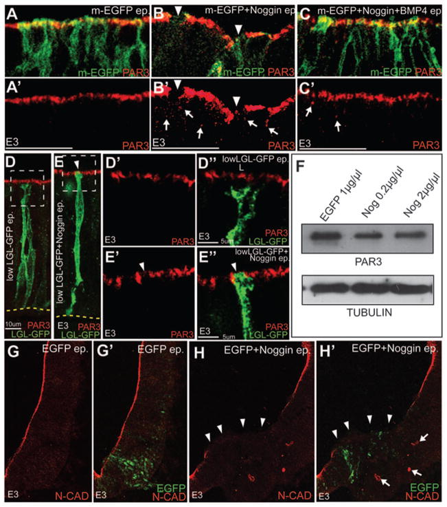
Figure 6. BMP signaling regulates epithelial apicobasal polarity.
(A–C′) Confocal images showing that the downregulation of PAR3 (red, arrowheads B, B′) labeling from the apical surface of Noggin + m-EGFP (B, B′) electroporated cells can be partially rescued by co-electroporation of Noggin+BMP4 (C-C′). Control (m-EGFP only) electroporations are shown in A, A′. A′, B′ and C′ show the PAR3 channels for the merges shown in A, B and C respectively. (D–E″) Control LGL-GFP electroporations showing the segregation of the apical (PAR3+) and basolateral (LGL+) compartments (D-D″). Co-electroporation of Noggin with LGL-GFP (1μg/μl) results in the apical removal of PAR3 and the apical incursion of LGL-GFP (E-E″). (F) Western blots showing that PAR3 levels are not different between control and Noggin-electroporated E3 whole-cell lysates (top row). Bottom Row: loading controls. (G–H′) Confocal images demonstrating that reduced N-CAD expression at the apical membrane (arrowheads in H, H′) and the ectopic cytoplasmic expression of N-CAD (arrows in H, H′) in Noggin-electroporated brains (i, arrowheads). Controls shown in G, G′.

