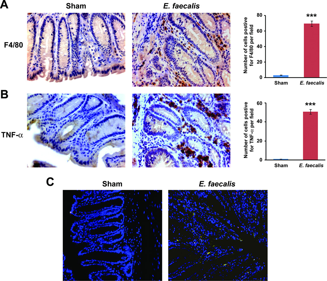
Figure 1. E. faecalis-infected macrophages produce TNF-α.
A Immunohistochemical staining of colons from Il10−/− mice colonized with E. faecalis for 9 months; increased numbers of macrophages (F4/80 positive, brown) and B, staining for TNF-α (brown) are noted in the lamina propria (40X), but not seen in shams. Numbers of F4/80 and TNF-α-positive cells per 20X field are increased in colons from Il10−/− mice colonized with E. faecalis for 9 months compared to sham (***P < 0.001). C Immunofluorescence staining of colon biopsies from E. faecalis colonized Il10−/− mice using F4/80 (red) and TNF-α (green) confirm co-localization of TNF-α to macrophages (yellow).

