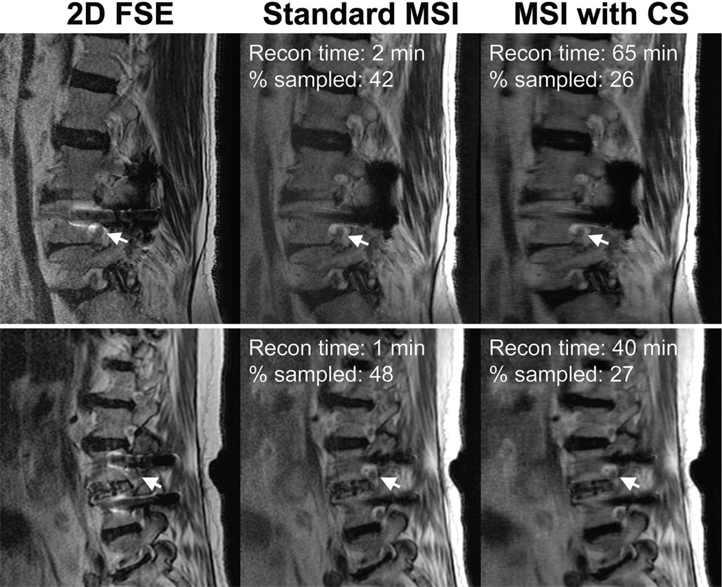
Figure 3.
Sagittal T2-weighted FSE and MSI slices from two subjects (top and bottom row) acquired at 1.5 T demonstrating the retrospective application of compressed sensing. 2D FSE images are shown to show the distortion induced by the presence of metal. The arrows point to nerves and neural foramina that are clearly depicted in the images acquired with MSI. The percentage sampled in the fully-sampled half-Fourier image is under 50% as corners in ky-kz space were not acquired.

