Fig. 5.

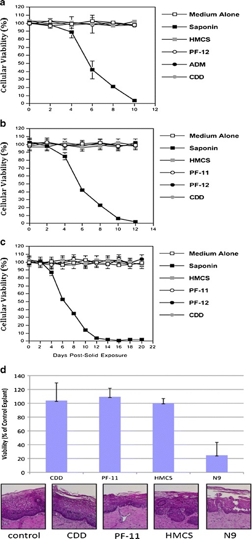
(a) Viability of human PBMC cells during 10 days exposure to HMCS, PF-12, ADM and CDD matrices. (b) Viability of cultured T-lymphocytes and (c) macrophages during 12 and 20 days exposures to HMCS, PF-11, PF-12, and CDD matrices. Data presented in (a), (b) and (c) are expressed in percentage of viable cells. The error bars in (a), (b) and (c) represent standard errors of duplicates. Data shown in panels (a), (b) and (c) are each representative of two independent experiments. (d) Ex vivo viability of human ectocervical tissue during 5 days exposure to CDD, PF-11, and HMCS matrices. The data represent the mean ± SD of three independent tissues performed in duplicate. The viability of the N9-treated tissues were significantly reduced from control (untreated) tissues (P < 0.05) (Wilcoxon T-test).
