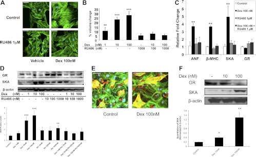
Fig. 1.
Glucocorticoids induce hypertrophy in rat cardiomyocytes. Rat H9C2 cells or isolated neonatal cardiomyocytes were treated for 72 h with Dex in the presence or absence of RU486. A, Representative confocal microscopic images of phalloidin-stained H9C2 cells. Scale bar, 45 μm. B, H9C2 cell volumes were measured by flow cytometry and are presented as percent volume change relative to the control cells. C, The mRNA levels in H9C2 cells for GR and the cardiac hypertrophic marker genes ANF, β-MHC, and SKA were determined by real-time PCR and normalized to cyclophilin B. D, GR and SKA protein levels in H9C2 cells were measured by immunoblotting. The lower panel shows quantification of SKA protein normalized to β-actin. E, Representative confocal microscopic images of primary cardiomyocytes double labeled with β-MHC (red) and phalloidin (green). The presence of actin and β-MHC in the same cell (yellow) distinguishes the cardiomyocytes from fibroblasts. Scale bar, 45 μm. F, GR and SKA protein levels in primary cardiomyocytes were measured by immunoblotting. The lower panel shows quantification of SKA protein normalized to β-actin. Data represent the mean ± sd from three independent experiments. *, P < 0.05; **, P < 0.01; ***, P < 0.001 vs. controls.

