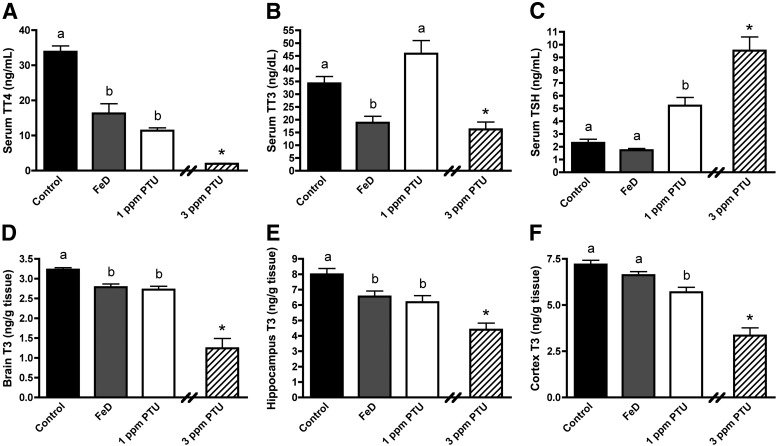
Fig. 1.
Fetal and neonatal Fe deficiency reduces serum and brain TH levels. A, Serum TT4. B, Serum TT3. C, Serum TSH. Serum was harvested from P10 male pups (n = 8–10). D, Whole-brain T3. E, Hippocampal T3. F, Cerebral cortical T3. Half brains (n = 6–9), hippocampi (n = 9–10), and cerebral cortices (n = 9) were harvested from P10 male pups. Data are presented as the mean ± sem. Groups not sharing a common superscript are significantly different by one-way ANOVA and Tukey's or Scheffé's multiple comparison test (P < 0.05). The 3-ppm PTU group served as a positive hypothyroid control. Asterisks indicate a statistical difference between 3 ppm PTU and control.

