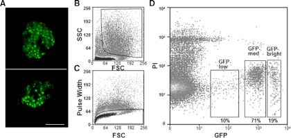
Fig. 1.
Three populations of pancreatic β-cells were identified from adult MIP-GFP mice. A, Fluorescence images of pancreatic islets isolated from adult MIP-GFP mice, observed with confocal microscopy. Scale bar, 100 μm. B, The first gate for pancreatic β-cells was set to analyze cell size and granule density estimated by FSC and SSC, respectively. C, The second gate employed pulse width and was used to exclude doublets or other cell clusters. D, The third gate was set for GFP and PI to exclude GFP-negative cells and dead cells.

