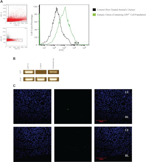
Fig. 1.
Uteri of endometriotic mice contain GFP+ cells from the ectopic lesions. A, FACS analysis of uterine tissue from mice with GFP+ endometriosis contains GFP+ cells. The two small panels on the left represent the gates for the sorted population of GFP+ cells: noncellular debris (upper panel) and single cells (lower panel). The large diagram represents the percentages of GFP+ cells (fluorescein isothiocyanate channel), in which the green plot represents endometriotic samples and the black plot the control. The linear segment labeled with an asterisk shows the percentage of GFP+ cells that were collected from the sample (1.8%). B, PCR confirms the expression of GFP in uteri collected from animals with GFP+ endometriosis. The GFP amplification product was observed only in the uteri of animals with endometriosis and not the control group. β-Actin was used as a loading control. C, Immunofluorescence was used to detect GFP protein expression in the mouse uterus. GFP expression was detected in stromal cells of the animals with GFP+ endometriosis and not in controls. In all the samples from the experimental group (n = 8), GFP-expressing cells were located in the basalis layer. BL, Basalis, LE, luminal epithelium. From left to right: GFP (green channel), 4′,6′-diamino-2-phenylindole (blue channel), and a merged channel. Scale bar, 100 μm.

