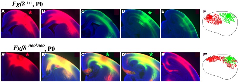
Figure 3.
Intra-neocortical projection patterns in P0 mice with rostral patterning defects (Fgf8neo/neo mutants) compared with P0 control littermates (Fgf8+/+). Hundred micrometer coronal sections presented in rostral to caudal series of brain hemispheres following DiI [red asterisk, (A,A′)] or DiA [green asterisk, (D,D′)] crystal placement the rostral and caudal neocortex (putative somatosensory and visual cortex, respectively), oriented with dorsal up and lateral to the right. Sections were analyzed for the distributions of retrogradely labeled cell bodies, with lateral view reconstructions shown in (F,F′). Hemi-sections from control mice (A–E) demonstrate no overlap of retrograde label from dye placements in putative somatosensory (A) or visual (D) cortex, as red and green label remain segregated. However, Fgf8neo/neo mutants showed a robust phenotype, indicated by red–green overlap [yellow label, (B′–D′)] and red label caudal to this overlap (E′) reflecting ectopic caudal projections to rostral somatosensory cortical locations. The ectopic intra-neocortical connections are easily observed in the reconstructions where caudal locations aberrantly project to rostral fields in the mutant (F′) but not in the control (F). (F,F′)-Rostral is left, dorsal up. Figure adapted from Huffman et al. (2004).

