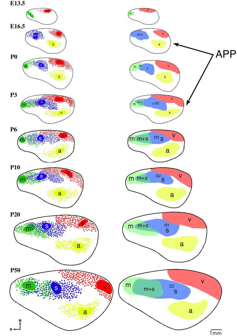
Figure 4.
Reconstruction of areal boundaries through analysis of intra-neocortical connections in E13.5-P50 wild-type mice. All panels represent a lateral view of one hemisphere. Left column: dye placement locations and organization of retrogradely labeled cells (colored patches, dye placement, and dye spread; red filled circles, retrogradely labeled cells in putative caudal/visual cortex; blue filled circles: retrogradely labeled cells in putative somatosensory cortex; green filled circles, retrogradely labeled cells in putative rostral/motor cortex; yellow filled circles, retrogradely labeled cells in putative auditory cortex; thick black line, hemisphere outline). Right column: lateral view reconstructions of putative areal boundaries as determined by INC analyses (colored areas, putative cortical areas as labeled; r, putative rostral area; c, putative caudal area; m, putative motor cortex; m + s, putative sensory-motor amalgam; m/s or s, putative motor/somatosensory or somatosensory cortex; a, putative auditory cortex; v, putative visual cortex). Areal patterning period (APP) is from E16.5-P3. Stars indicate location of putative barrel field. Oriented medial (M) up and rostral (R) to the left. Scale bar = 1 mm. Adapted from Dye et al. (2011a,b).

