Figure 1 .
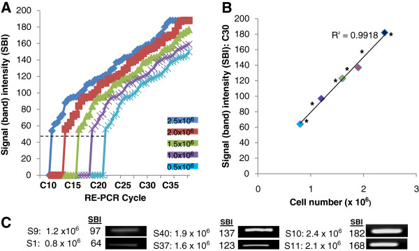
RE-PCR using DNA from HGF-1 cells (standards) and saliva samples. A) DNA standards obtained from HGF-1 cells (0.5 – 2.5 x 10⁶ cells/mL) established minimum threshold (CT) and saturation (CS) cycles; (high cell concentration) 2.5 x 10⁶ cells/mL CT = C10, CS = C35; (low cell concentration) 0.5 x 10⁶ cells/mL , CT = C20, CS = C45. B) RE-PCR at C30 (above low concentration CT = C20, below high concentration CS = C35) revealed strong, positive correlations (R2 = 0.9918) between signal band intensity (SBI) and cell concentration. C) RE-PCR using DNA extractions from all saliva samples produced bands with increasing SBI; two representative saliva samples with low (0.8 – 1.2 x 10⁶ cells/mL), mid (1.6 – 1.9 x 10⁶ cells/mL) and high (2.1 – 2.4 x 10⁶ cells/mL) cell concentrations are shown. Plotting the sample SBI (*) with the DNA standards revealed near perfect alignment.

