
Figure 9. Evidence for ATP release from type II cells following activation of P2Y2 receptors using petrosal neurones as ATP biosensors.
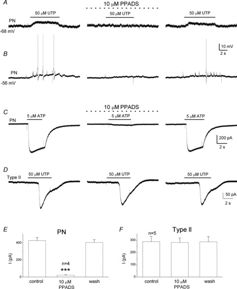
Using the co-culture configuration described in Fig. 8, application of UTP over the tripartite complex evoked depolarization (A) or action potential firing (B) in the juxtaposed PNs. In both cases the response was reversibly abolished during perfusion of the P2X2/3 receptor blocker PPADS (10 μm; middle traces), consistent with release of ATP. Note that 10 μm PPADS almost completely abolishes ATP-evoked currents in isolated PNs (C and E; for E, ***P < 0.001, ANOVA), but fails to affect UTP-induced whole-cell currents in type II cells (D and F; for F, P > 0.05, ANOVA).